
Modern medical education requires accurate anatomical representations to bridge the gap between theory and practice. While digital visualization tools have advanced rapidly, medical students still require physical, tactile feedback to develop critical spatial memory. DIGIHUMAN addresses this challenge by converting high-precision digital datasets into durable physical assets. This technology provides laboratories with a sustainable, accurate alternative to traditional specimen preservation.
The Foundation of Precise Data Extraction
Manufacturing high-fidelity medical models begins with reliable source data. The production process extracts information from comprehensive digital human datasets. These sets include refined segmentation data and original sectional imaging.
To achieve microscopic accuracy, the DIGIHUMAN software utilizes a voxel size of 0.0384 mm× 0.0384 mm× 0.1 mm. Engineers map voxels directly from the surface of each anatomical structure to generate an exact texture map. This extraction process helps maintain geometric morphology that closely matches human anatomical structures without unnecessary artistic interpretation. Technicians verify this by comparing digital outputs against traditional formalin-fixed cadavers to ensure strict structural consistency.
The Industrial Multi-Channel Printing Process
Translating these massive volumetric datasets into physical models requires specialized industrial hardware. Standard desktop printers cannot replicate the structural complexity of human tissue. Advanced manufacturing facilities utilize drop-on-demand digital inkjet technology combined with rapid light-curing systems.
These industrial printers operate using 12 distinct material channels simultaneously. The printheads feature up to 3,840 piezoelectric spray holes. During production, the hardware deposits microscopic droplets of liquid resin layer by layer. An integrated ultraviolet light source immediately cures each layer upon deposition. Through this high-frequency spray curing, production volume reaches 4 liters per hour.
This immediate curing process significantly reduces post-processing. Furthermore, the system automatically generates dissolvable support structures. Depending on the printing material, temporary support structures can be removed through appropriate post-processing methods. This helps ensure that delicate, overhanging geometries and microscopic neural branches remain neatly intact during the high-speed build phase.
Advanced Material Integration and Properties
Human anatomy consists of vastly different physical densities. A high-quality printed model must accurately reflect these varying mechanical properties. To achieve this, the 12-channel system utilizes a diverse spectrum of specially formulated, environmentally friendly resins.
The hardware seamlessly blends rigid polymers with elastomeric rubber-like materials in a single automated run. The system also introduces full-color CMYK mixing alongside white and transparent resins. This multi-material capability allows manufacturers to create composite anatomical structures.
For example, a printed model can feature a soft, flexible liver structure housing rigid internal bile ducts, all encased in a transparent outer shell. This allows students to physically handle the organ while observing its internal spatial relationships. The resulting tactile feedback provides a more realistic hands-on learning experience than digital visualization alone. Unlike real tissue or wet specimens, these photo-cured resins are highly durable, resisting environmental degradation, moisture changes, and wear from repeated student handling.
Specific Applications in Topographical Anatomy
The primary application of these industrial prints is the modernization of university anatomy laboratories. Certain anatomical systems are notoriously difficult to dissect and preserve in traditional wet labs. 3D printing provides a permanent solution for studying these complex structures.
In neuroanatomy, the printing process accurately reproduces the intricate branching of the cranial nerves and the central nervous system. In angiology, microscopic blood vessels that typically degrade in biological specimens are printed using vibrant, color-coded resins. This high-contrast coloration makes arterial and venous pathways instantly identifiable.
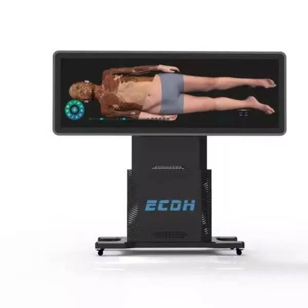
These physical models serve as a vital counterpart to digital instruction. While an interactive anatomy table excels in virtual dissection and deep visual exploration, these printed replicas build the necessary physical hands-on skills. Together, they create a comprehensive learning environment that covers both digital visualization and tactile engagement.
Standardizing Resources for Laboratory Curricula
Medical universities face recurring costs and biological hazards associated with traditional wet labs. Transitioning to high-precision 3D printing models resolves these logistical burdens.
Because production relies on automated digital extraction, every manufactured piece maintains strict structural integrity. Educators can select specific color configuration schemes to highlight anatomical systems. This makes complex networks, such as the cranial nerves or cardiovascular paths, instantly identifiable.
The printed model maintains high geometric consistency with the original digital dataset, although minor manufacturing variations may occur. This standardization helps provide consistent practical learning resources for different student cohorts. It also provides a safe, chemical-free alternative to traditional lab materials.
Applications in Surgical Preparation and Consultation
The utility of these 3D-printed replicas extends to professional clinical environments. Surgeons rely on exact structural representations for preoperative spatial preparation.
A patient-specific model allows a surgical team to evaluate the physical relationship between a tumor and surrounding critical blood vessels before entering the operating room. This may help surgeons better understand anatomical relationships before surgery.
These physical tools also bridge the communication gap in doctor-patient consultations. Medical terminology and radiological imaging often confuse patients. By physically holding a model of their anatomy, patients gain a clearer understanding of their diagnosis. This transparency can help build trust and contribute to improved patient outcomes. By integrating high-fidelity manufacturing, DIGIHUMAN supports both education and clinical practice.