
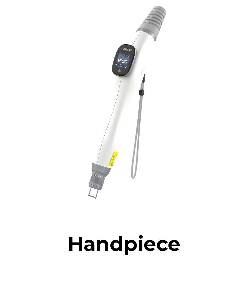
Accurate assessment of pigment depth remains a primary determinant in structuring safe and effective laser removal protocols. Utilizing a high-output Q switched ND YAG laser machine allows clinicians to target tattoo pigments at different dermal depths while managing energy delivery according to treatment requirement. Engineering innovations from manufacturers such as ENZOEYS emphasize optical precision and sustained energy output, enabling practitioners to address complex, layered tattoo pigments systematically.
Understanding how ink particles distribute within cutaneous tissue requires evaluating both professional application techniques and biological pigment migration over time. Through careful physical examination and light-tissue interaction principles, operators can tailor fluence settings to match specific ink depths.
Anatomical Distribution of Trapped Dermal Inks
Tattoo placement alters natural dermal structures by depositing insoluble metallic or organic pigments into the papillary and reticular dermis. Professional tattoos typically place ink deeper and with greater density compared to amateur applications, which exhibit irregular, superficial distributions.
Over time, dermal macrophages engulf pigment particles, trapping them within fibrous tissue matrixes at varying levels. Evaluating the age and texture of a tattoo can provide additional context when assessing whether pigment is likely to be distributed more superficially or deeper within the dermis.
Light Absorption Physics Across Dermal Depths
Optical penetration depth correlates directly with the wavelength emitted by short-pulse lasers. Shorter wavelengths scatter more rapidly in superficial tissue, whereas longer wavelengths penetrate deeply into the reticular dermis to reach encapsulated ink deposits.
Deploying a calibrated Q switch ND YAG laser machine enables operators to utilize distinct wavelengths tailored to pigment depth and color. Wavelengths such as 1064nm penetrate deeply into dermal structures, targeting dark, deeply placed inks while minimizing superficial epidermal absorption.
Diagnostic Visual and Palpation Assessment Techniques
Before initiating optical exposure, clinicians perform tactile and visual evaluations to identify tissue changes associated with deep ink placement. Raised or hypertrophic areas often indicate heavy pigment loads or pre-existing scarring from original tattooing procedures.
Dermoscopy and oblique lighting assist practitioners in evaluating surface ink density and subtle color variations. Identifying underlying scarring helps technicians adjust pulse energy and spot sizes to prevent exacerbating existing dermal trauma.
Photomechanical Fragmentation and Particle Size Reduction
High-peak power short pulses generate photoacoustic shockwaves that shatter dense ink clusters into microscopic fragments. Rapid mechanical stress fractures locked pigment deposits, allowing local phagocytes to transport smaller ink debris through lymphatic channels.
Sustained thermal stability becomes critical when treating high-density, deeply placed inks during lengthy clinical procedures. Systems engineered like the LIFFAN Q6 support extended 24-hour continuous operation, maintaining steady pulse output across demanding treatment schedules without energy attenuation.
Strategic Parameter Selection for Variable Pigment Layers
Effective treatment strategy requires adjusting fluence, spot size, and repetition rates based on estimated pigment depth. Initial sessions typically focus on superficial ink layers using moderate fluences to clear upper dermal density safely.
Subsequent sessions can be adjusted according to the remaining pigment depth, with spot size and fluence selected based on the tattoo characteristics and treatment response. Gradual treatment across multiple sessions allows fragmented pigment to be progressively cleared between appointments.
Clinical Versatility Across Multi-Indication Workflows
Modern clinical practices require equipment capable of handling broad dermatological portfolios beyond tattoo removal alone. High-performance platforms adapt to diverse skin conditions through versatile pulse modalities and adjustable energy parameters.
Integrating multi-functional systems like the LIFFAN Q6 offers comprehensive treatment coverage for benign pigmentation, acne, and skin rejuvenation. Broad clinical adaptability maximizes operational efficiency, allowing facilities to address various client needs with a single versatile device.
Tissue Recovery Protocols and Lymphatic Clearance Rates
Post-treatment healing phases depend heavily on the depth of optical disruption and individual lymphatic system efficiency. Thermal stress within deeper dermal layers requires adequate recovery intervals to prevent secondary hyperpigmentation or scarring.
Spacing treatment sessions 6 to 8 weeks apart allows macrophages sufficient time to clear fragmented ink particles. Proper post-care guidelines, including sun avoidance and barrier repair, support natural dermal recovery and optimize long-term clearing results.
Clinicians should also assess individual lymphatic function and inflammatory response patterns during initial consultations, as patients with compromised clearance mechanisms may require extended intervals between sessions or modified energy settings to avoid overtreatment. Documenting recovery trajectories across successive visits enables practitioners to refine protocol timing for each patient—transforming standard schedules into personalized recovery plans that balance clinical efficacy with tissue safety.
Equipment Reliability and Long-Term Performance Metrics
Selecting optical hardware for high-volume practices involves evaluating component durability and thermal dissipation capabilities under continuous use. Inconsistent power output during treatment sessions leads to uneven pigment fragmentation and unpredictable clinical timelines.
The Q6 is designed for 24-hour continuous operation, supporting facilities with demanding daily treatment schedules. Reliability in energy delivery directly protects patient safety and ensures reproducible clinical outcomes over extended operational cycles.
Conclusion
Mastering ink depth assessment enables clinicians to design targeted, multi-session clearance protocols tailored to individual dermal structures. Combining wavelength selectivity with stable nanosecond energy delivery minimizes tissue trauma while optimizing particle fragmentation. Equipment engineered by ENZOEYS demonstrates how high-capacity systems support daily practice workflows, allowing facilities to deliver safe, effective treatments across diverse dermatological and tattoo removal indications.